OCT-A for Acute Stroke
This Webapp allows to estimate the likelihood of acute stroke using the as input vessel density variables from OCT-A. It has been developed at the Giancardo lab at UTHealth as part of an effort to develop systems and data for identifying acute strokes using retina imaging. This would enable the deployment of life-saving drugs in situations where brain scans are typically not available such as ambulances or even space missions.
Input
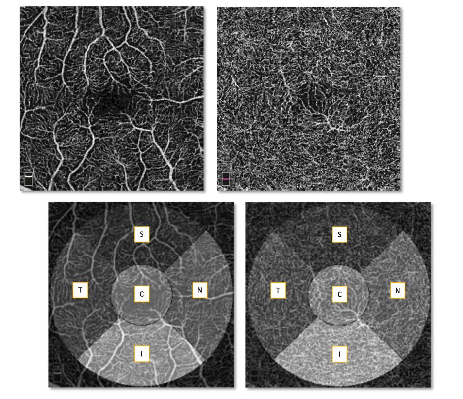
The algorithm requires 10 variables per retina estimated with an Optovue Avanti OCT-A Camera and the iVue software. OCT-A en-face images with superficial (identified as L1) and deep (identified as L2) layers needs to be used. More information is available in this publication which should also be used as citation: S. Pachade, I. Coronado, R. Abdelkhaleq, J. Yan, S. Salazar-Marioni, A. Jagolino, M. Bahrainian, R. Channa, S. A. Sheth, L. Giancardo. "Detection of Stroke with Retinal Microvascular Density and Self-Supervised Learning Using OCT-A and Fundus Imaging " J. Clin. Med. 2022, 11(24), 7408; https://doi.org/10.3390/jcm11247408
Note that the algorithm has been trained on a limited dataset, and that the scores generated are for research purposes only, not for clinical care. See the disclaimer in the bottom of the page.